
Utilization of Phase Contrast Magnetic Resonance Imaging to Measure Cerebral Hemodynamic Changes Before and After a NUCCA Atlas Correction – A Case Series
Results of a case study describing a subject diagnosed with migraine headache (without aura) reveal compelling results obtained with Phase Contrast Magnetic Resonance Imaging Angiography (PC MR). The subject was evaluated using the protocol of the National Upper Cervical Chiropractic Association (NUCCA) to determine the presence of an Atlas misalignment. After Atlas correction, a follow up PC MR Study demonstrated changes in cerebral venous outflow. A change in vessel outflow pattern from a jugular to the paravertebral plexus route was discerned. Venous flow rate and vessel pulsatility decreased as well as cerebrospinal flow rate across the Atlas (C-1) vertebra. Most significantly, the imaging procedure measured a distinct decrease in intracranial compliance. The subject obtained relief from migraine headache pain consistent to maintenance of his Atlas correction by the end of thirty days. The PC MR exam continued to show improvement of the hemodynamic parameters measured over the 16-week study period consistent to maintenance of Atlas alignment.
These results coupled with previously documented normalization of blood pressure in a randomized double blind study indicate a casual physiologic and measureable effect occurs after the correction of an Atlas misalignment. Further investigation facilitates elucidation of a physiologic mechanism subsequent to misalignment and its correction. The potential resulting from this study in alleviating migraine pain resultant to an Atlas correction may provide evidence of a viable alternative to usual and customary treatment. A case series, imaging ten subjects diagnosed with migraine headache by a Neurologist is necessary to demonstrate consistency in the cerebral hemodynamic effects previously observed.
SPECIFIC AIMS
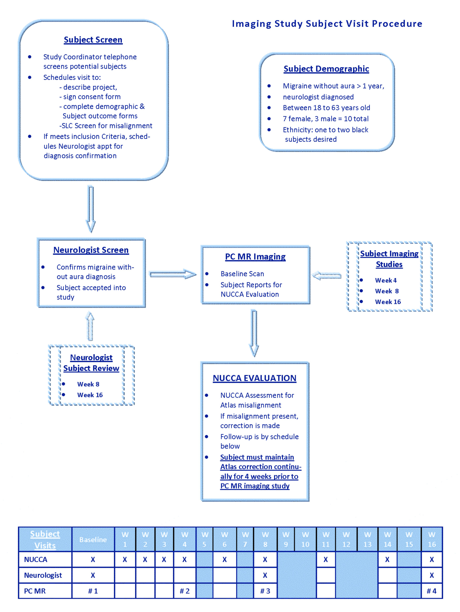
- Document the effect of Atlas correction on Cerebral Venous and CSF outflow patterns and velocities, with decreased intracranial compliance, as measured by PC MRA in a case series study of ten migraine (without aura) neurologist diagnosed subjects.
- Observe similarities and differences in a subject’s subjective response to correction and ablation of migraine symptoms, headache pain, in relation to documented changes in PC MRA.
- Demonstrate PC MR measured results and velocities are reproducible, sustained, and consistent over time, after an Atlas correction.
Possible Atlas misalignment interaction with the Trigeminovascular System via decreased intracranial compliance may produce neural influences on cerebral circulation as proposed by Dr. Goadsby providing impetus for further study. This study is significant in bringing a new imaging technology into Canada while offering another research facility to conduct productive investigation in determining a physiologic mechanism underlying the Atlas misalignment and its correction. A case series of ten migraine subjects monitored by PC MR measures cerebral circulatory response to the Atlas correction with a potential to reveal much new information of an underlying physiologic mechanism.